Free cancellation up to 2 weeks before the course starts, installment payment possible with down payment
Free places: 4
RegisterUniklinik RWTH Aachen
Pauwelsstraße 30
52074 Aachen
Wed, 26. Aug. 2026: 15:00-20:45
Thu, 27. Aug. 2026: 15:00-20:45
Fri, 28. Aug. 2026: 15:00-20:45
Sat, 29. Aug. 2026: 10:30-16:15
For questions: simcenter@schallware.de, Telephone +491774911854
 ⯈
⯈
Euregional Advanced Abdominal Ultrasound, Aachen/Aken/Aix-la-Chapelle
EFSUMB DEGUM KBV-compliant, simulation-supported with integrated pathology database including us-scanning of volunteers including IBD
https://www.schallware.de/rental/2759
Euregional Advanced Abdominal Ultrasound, Aachen/Aken/Aix-la-Chapelle
Scientific Directors:
• Dr. Karim Hamesch, Gastroenterologist/Internist, University Hospital Aachen, Germany
• Dr. Robert de Knegt, Gastroenterologist/Hepatologist, Erasmus MC University Medical Center, Rotterdam, the Netherlands
• OFA Dr. med. Michael Ludwig DEGUM 3, Berlin
Lecturers and Tutors:
• Dr. Jamal Ali, Gastroenterologist/Internist, University Hospital Cologne
• Dr. Susanne Fleig, Internist/Nephrologist, University Hospital Aachen, Germany
• Prof. Dr. Christian Jenssen, Hospital Märkische Oberland, Strausberg, Germany
• Dr. Claudia Lucius, Gastroenterologist/Internist, Helios Clinic Berlin-Buch, Germany
• Dr. Michael Ludwig, Internist/Pulmonary Medicine, Innere Bundeswehrkrankenhaus Berlin, Germany
• Prof. Alina Popescu, Gastroenterologist, Victor Babes University of Medicine and Pharmacy, Timisoara, Romania
• Dr. Matteo Rosselli, University College London, London, UK
- Dr. Bart Takkenberg, Gastroenterologist/Hepatologist, Amsterdam UMC, Amsterdam, the Netherlands
• Prof. Roxana Sirli, Gastroenterologist, Victor Babes University of Medicine and Pharmacy, Timisoara, Romania
• Dr. Pavel Taimr, Gastroenterologist/Hepatologist, University Hospital Prague, IKEM Praha, Czech Republic
Ultrasound devices: 1 US device per 5 participants with one healthy volunteer
Schallware Ultrasound Simulators: Advanced 1 participant per simulator + master simulator on stage
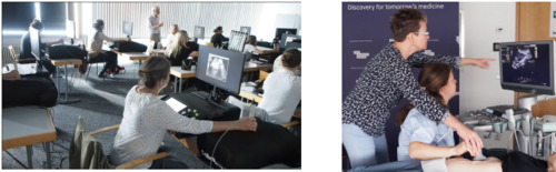
These simulation-supported ultrasound courses last 4 days. The courses are conducted with alternating lecturers and tutors on real US devices and simulators.
The aim of the courses is to offer a basic and advanced abdominal course according to EFSUMB/DEGUM guidelines. Participants will have extensive individual practice time using the simulator and gain access to the pathology database.
The lecturers perform a “master scan” on stage with projection via beamer, while all participants repeat the exercise simultaneously using a dummy probe and mannequin (simulator).
After simulator training, the acquired skills are applied to volunteers and real ultrasound devices.
Contents of the courses:
• Preparation: short online instructional videos, mandatory prior to the course (Methodology and vascular anatomy, approx. 15 min each).
Links are sent in advance during registration.
• Interactive short lectures
• Live demonstrations on the ultrasound devices (incl. knobology, artifacts, Doppler, thorax, bowel, liver)
• Practical exercises on the ultrasound device: guided by tutors and lecturers, 15 teaching units (simulators), 5 participants per regular US device.
• Simulator practice sessions: topic-related, directly after the theoretical introduction in the short lecture, or moderated during the simultaneous master scan.
Lecturers and tutors introduce the respective organs or organ systems with short lectures. Afterwards, participants independently work out normal findings and typical pathological findings using real patient case studies. Participants use the simulators, where case studies (clinical data and virtual models) can be uploaded.
Lectures are supported by examinations on a patient dummy, into which real three-dimensional patient data are virtually projected.
Sonographic case studies on the simulator with real patient data examples:
• Normal findings of all presented organs and organ systems
• Aortic aneurysm
• Aortic sclerosis
• Pancreatic lipomatosis
• Pancreatitis
• Pancreatic carcinoma
• Hydronephrosis
• Nephrolithiasis
• Renal cysts
• Renal tumors
• Fatty liver
• Liver cysts
• Liver tumors
• Liver cirrhosis, portal hypertension
• Bile duct dilatation
• Gallstones (cholecystolithiasis and choledocholithiasis)
• Cholecystitis
• Splenomegaly
• Ascites
Language during the courses: English
Each topic: ultrasound simulators.
Live scan with volunteers/patients when indicated.
Advanced ultrasound course Abdomen and Retroperitoneum (incl. Kidneys, IBD), Thorax (without Heart)
Wednesday, 26.08.26: Tag 1
Thursday, 27.08.26: Tag 2
Friday, 28.08.26: Tag 3
Saturday, 29.08.26: Tag 4
|
Red bars show acquired areas of patient. Red arrows equate to intercostal fan volumes.
Cases were acquired for example with convex, linear probe, in color doppler modus and maybe at several dates. You can open details and browse image list of regions of interest (ROIs), which are supported by simulator as you can be led automatically to these structures (bubble in space).
| Name | Location | Keywords | Technology | Probe | |
|---|---|---|---|---|---|
| Patient 126 |  | Acute appendicitis | convex, linear | Details | |
| Patient 2 |  | Hydatid cyst WHO CE3a | convex | Details | |
| Patient 20 |  | 60 y/o male, Anatomy | convex | Details | |
| Patient 28 |  | Liver metastases, Neuroendocrine tumors NET (unclear origin) | convex | Details | |
| Patient 31 |  | Horse shoe kidney, Abdominal mass, Arteriosclerosis | convex | Details | |
| Patient 41 |  | Acute splenic rupture and necrotizing pancreatitis (tail), intraperitoneal bleeding | convex | Details | |
| Patient 53 |  | Caval thrombus, renal cell carcinoma RCC (left), liver metastases | convex | Details | |
| Patient 54 |  | Tumor infiltration of the kidney, obstructive uropathy | convex | Details | |
| Patient 59 |  | FNH in S5 | convex | Details | |
| Patient 64 |  | Acute pancreatitis, choledocholithiasis, cholecystolithiasis, liver cyst | convex | Details | |
| Patient 65 |  | Aneurysm and septic thrombosis of portal and splenic vein | convex | Details | |
| Patient 71 |  | Infected splenic cyst | convex | Details | |
| Patient 77 |  | Mechanical small bowel ileus | convex | Details | |
| Patient 89 |  | 60 y/o female, Urothelial carcinoma | convex, color | Details | |
| Patient 91 |  | Severe steatohepatitis, Asc.-Decompensation, HCC | convex | Details | |
| Patient 92 |  | Acute cholecystitis with lithiasis, detritus, fatty liver | convex | Details | |
| Patient 96 |  | LTX, CBD with stone sludge, ITBL, thrombus in hepatic artery stump | convex | Details | |
| Patient 97 |  | Pancreatic pseudocyst, cholecystolithiasis, hydronephrosis, right ureteral occlusion, parapelvic cysts | convex | Details | |
| Patient 98 |  | Marfan syndrome, HTX, aortic aneurysm, aortic dissection | convex, color | Details | |
| Patient 100 |  | DLT, NHL Liver, infiltrated intestine, bilateral pleural effusion, gallbladder wall edema (hypalbuminemia) | convex | Details | |
| Patient 101 |  | Peritoneal carcinosis by liposarcoma | convex | Details | |
| Patient 103 |  | Liver hemangiomas, cholecystolithiasis | convex | Details | |
| Patient 105 |  | Appendicitis with fecalith - atypical course | linear | Details | |
| Patient 111 |  | Primary sclerosing cholangitis PSC | convex | Details | |
| Patient 115 |  | Right pleural effusion | convex | Details | |
| Patient 116 |  | Left pleural effusion | convex | Details | |
| Patient 117 |  | Liver cirrh., reopened umbilical vein, gallbladder hydrops, cholecystolithiasis | convex | Details | |
| Patient 118 |  | Liver cysts, M.Crohn, Adenomyomatosis, cholecystolith., nephrolith., stenosis of ileoc. anastomosis | convex, linear | Details | |
| Patient 120 |  | Hepaticojejunostomy, aerobilia, renal cyst, pancreatic pseudo cyst | convex | Details | |
| Patient 124 |  | Hepatic hemangioma, cholecystolithiasis, dilated CBD, ureteral stenosis | convex | Details | |
| Patient 125 |  | Free fluid, renal cysts, arteriosclerosis | convex | Details | |
| Patient 126 |  | Acute appendicitis | convex, linear | Details | |
| Patient 127 |  | Thoraco-Abdominal aortic aneurysm, right pleural effusion | convex, color | Details | |
| Patient 129 |  | Liver cirrhosis, nodules, cavathrombus, spleen with scar, left pleural effusion | convex | Details | |
| Patient 131 |  | Infrarenal aortic aneurysm | convex, color | Details | |
| Patient 132 |  | Bright liver tumors, splenomegaly | convex | Details | |
| Patient 133 |  | Polycystic liver and kidneys | convex | Details | |
| Patient 134 |  | Cholecystolithiasis with multiple concrements | convex | Details | |
| Patient 136 |  | Lymphomata in hepatoduodenal ligament and in splenic hilum | convex | Details | |
| Patient 141 |  | Aerobilia, Choledocholithiasis | convex | Details | |
| Patient 143 |  | Budd-Chiari and Cruveilhier-Baumgarten syndromes, Ascites (color) | convex, color | Details | |
| Patient 145 |  | Adrenal tumor, CEUS | convex, color | Details | |
| Patient 147 |  | Psoas abscess, Cholecystolithiasis, chronic cholecystitis | convex, linear | Details | |
| Patient 148 |  | Liver metastasis, cholecystolithiasis, chronic cholecystitis, melanoma | convex | Details | |
| Patient 149 |  | Portal vein thrombosis, TIPS, hypertrophic additional spleen | convex | Details | |
| Patient 150 |  | Liver tumors, CBD stenosis, enlarged lymph nodes | convex | Details | |
| Patient 151 |  | Hepatic Echinococcus alveolaris, dilated hepatici, purulent peritonitis | convex | Details | |
| Patient 153 |  | Liver cirrhosis, portal hypertension, cholecystolithiasis, partial portal vein thrombosis | convex | Details | |
| Patient 157 |  | Thrombosis of V.cava, Aneurysm | convex, linear, color | Details | |
| Patient 158 |  | Liver metastasis, Gallbladder cancer, Lymph node metastasis | linear, color | Details | |
| Patient 159 |  | Stenotic tumor of Asc.colon, Lymph node metastasis | convex, linear, color | Details | |
| Patient 22 |  | Ulcerative colitis treatment, anti-TNF-α | convex, linear, color | Details | |
| Patient 23 |  | Morbus Crohn, ileitis, segmental colitis, lymphadenitis | convex, linear | Details | |
| Patient 21 |  | M.Crohn, Distal sigmoiditis, Ascites, Lymph node enlargement, Sigmoidal hyperemia | convex, linear, color | Details | |
| Patient 14 |  | M.Crohn, Liver cysts, Adenomyomatosis, Cholecystolithiasis, Nephrolithiasis, Stenosis of ileocecal anastomosis | convex, linear | Details | |
| Patient 11 |  | Terminal ileitis Crohn, enterocutaneous fistula, psoas abscess | convex, linear, color | Details | |
| Patient 10 |  | Crohn's disease (sigmoid), Sigmoid stenosis, involvement of cecum-colon-ileum | convex, linear, color | Details | |
| Patient 9 |  | Colon carcinoma at right flexure, Paracolic abscess and malignant infiltration | convex | Details | |
| Patient 8 |  | Infiltrated appendix | convex, linear, color | Details | |
| Patient 6 |  | Crohn's disease relapse, Stenosis in neoterminal ileum with large fistula | convex, color | Details | |
| Patient 5 |  | Liver cysts, Crohn's disease, Stenosis of ileocecal anastomosis | convex, linear, color | Details | |
| Patient 4 |  | Terminal ileum with stenosises and interloop abscesses | convex | Details | |
| Patient 1 |  | Normal terminal ileum and appendix | linear | Details |
After registration you will receive a confirmation email with invoice and the course program. Payment on account or at any time with PayPal.
×Joining the waitlist requires a deposit of 250.00 €. As soon as a spot opens up you move up and the deposit is credited towards the course fee.
×